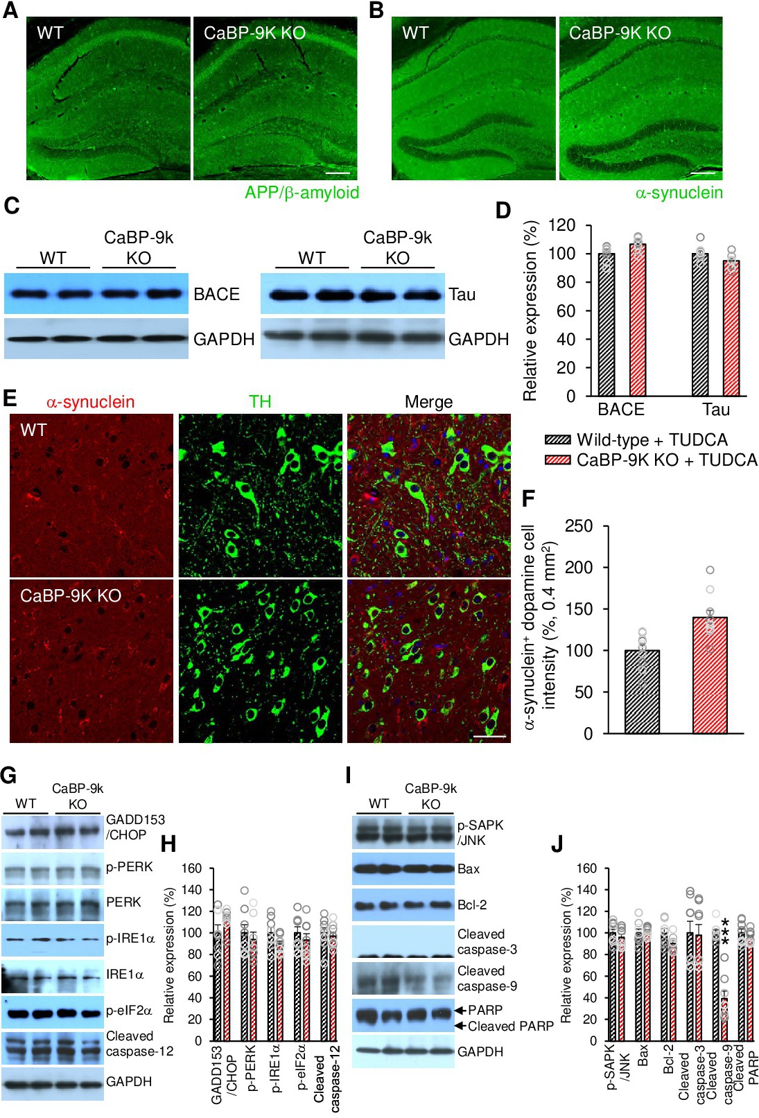
Fig. 6. TUDCA inhibits ER stress-induced apoptosis in CaBP-9k KO mice. (A) Immunofluorescence for APP/β-amyloid plaques in hippocampi of old TUDCA-treated wild-type and CaBP-9k KO mice. Scale bar= 200 µm. n = 4 mice for each group. (B) Immunofluorescence for α-synuclein in hippocampi of old TUDCA-treated wild-type and CaBP-9k KO mice. Scale bar= 200 µm. (C) Western blotting for BACE and Tau expression in brain lysates from TUDCA-treated wild-type and CaBP-9k KO mice. (D) Quantification of C. n = 8 mice for each group. (E) Immunofluorescence staining for α-synuclein and TH in dopaminergic cells in the SNc and VTA in old TUDCA-treated wild-type and CaBP-9k KO mice. Scale bar= 40 µm. (F) Quantification of E. n = 4 for mice for each group. (G,I) Western blotting for ER stress- and apoptosis-related proteins in old TUDCA-treated wild-type and CaBP-9k KO mice. (H,J) Quantification of G and I. n = 4 mice for each group. The intensities of the protein bands were normalized to the GAPDH level. Data shown are the means ± SEMs and were analysed by two-tailed unpaired Student's t-tests.